


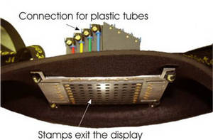
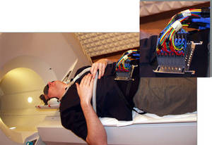
Functional Magnetic Resonance Imaging (fMRI) is a method of brain imaging. Briefly, a local measurement of the oxygen consumption gives information about activities in certain areas of the cortex (BOLD effect). The spatial resolution of fMRI measurements amounts to a few millimeters. Applying an ultrafast fMRI method known as 'echo-planar imaging' (EPI) acquires one image of the brain (containing more then 20 slices) in 2 seconds. The measurements take place in a strong static magnetic field (~2 Tesla) with an alternating electromagnetic field superimposed on it. It is therefore difficult to supply sensory stimulation. The operation of electromechanical and electronic systems is not possible in a magnetic field of this strength as such systems would also influence image acquisition. The Heidelberg group has developed an especially designed pneumatic tactile display (PTD), which can project tactile patterns on the skin of a proband. The patterns are generated with a dedicated software which is easy to control. The PTD has a modular structure so that different surface can be set up. Each module consists of 4 taxel (tactile elements).
Properties of the PTD:
The actual studies aim in the exploration of brainplastisity by sensory substitution. For this aim a studies with blind subjects and a sighted control group are carry out. The paradigms are designed to presented "visual like" tactile stimuli with the PTD. Three fMRI enquiries should demonstrate the variances in perception during training.
To compare the results with the above study and for a deeper matter in the kind of perception with the PTD a study with 12 native sighted subject is carry out. During this event-related fMRI experiment the subject has to distinguish between patten with differen properties. Both study are done in the framework of the SenSub-project.


One finished experiment focus on the examination of the shape of the tactile hemodynamic response in the human brain. Furthermore, a second experiment probes for dependencies on vibratory tactile stimulation in the frequency range of 2 to 6Hz.
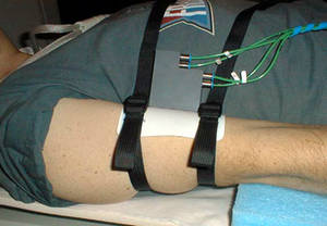
In previous experiments the activation induced from stimulation of index finger, arm and foot has been localized in the primary and secondary somatosensory cortex. Mapping of the somatotopic representation in the cerebral cortex which is called sensory homunculus has been performed in both primary and secondary cortex. For these investigations a smaller version of the PTD, consisting of one module (4 taxels), is mounted on the appropriate spot of the proband. We chose a walking pattern which alternates with rest phase. Each phase consists of 10 images and takes 40 seconds (one image of the brain takes here 4 seconds). All in all 200 images are recorded for each experiment.

 HOME
HOME
